Dental Pad Diagram . there are five teeth in each quadrant, composed of two incisors (central and lateral), a canine, and two molars. use our tooth chart template specifically designed for dentists. the present study provides detailed anatomical description of the parotid and mandibular salivary glands of the one humped camel with their. a free dental library of interactive 3d models for dental education including dental anatomy, occlusion, prosthodontics and endodontics. the pad contains glandular tissue, loose areolar connective tissue, the lower margin of the pterygomandibular raphe, fibers of the buccinator,. tooth diagrams, numbering systems, and color coding. Tooth diagrams for recording dental conditions are available with a.
from stock.adobe.com
Tooth diagrams for recording dental conditions are available with a. use our tooth chart template specifically designed for dentists. there are five teeth in each quadrant, composed of two incisors (central and lateral), a canine, and two molars. tooth diagrams, numbering systems, and color coding. the present study provides detailed anatomical description of the parotid and mandibular salivary glands of the one humped camel with their. the pad contains glandular tissue, loose areolar connective tissue, the lower margin of the pterygomandibular raphe, fibers of the buccinator,. a free dental library of interactive 3d models for dental education including dental anatomy, occlusion, prosthodontics and endodontics.
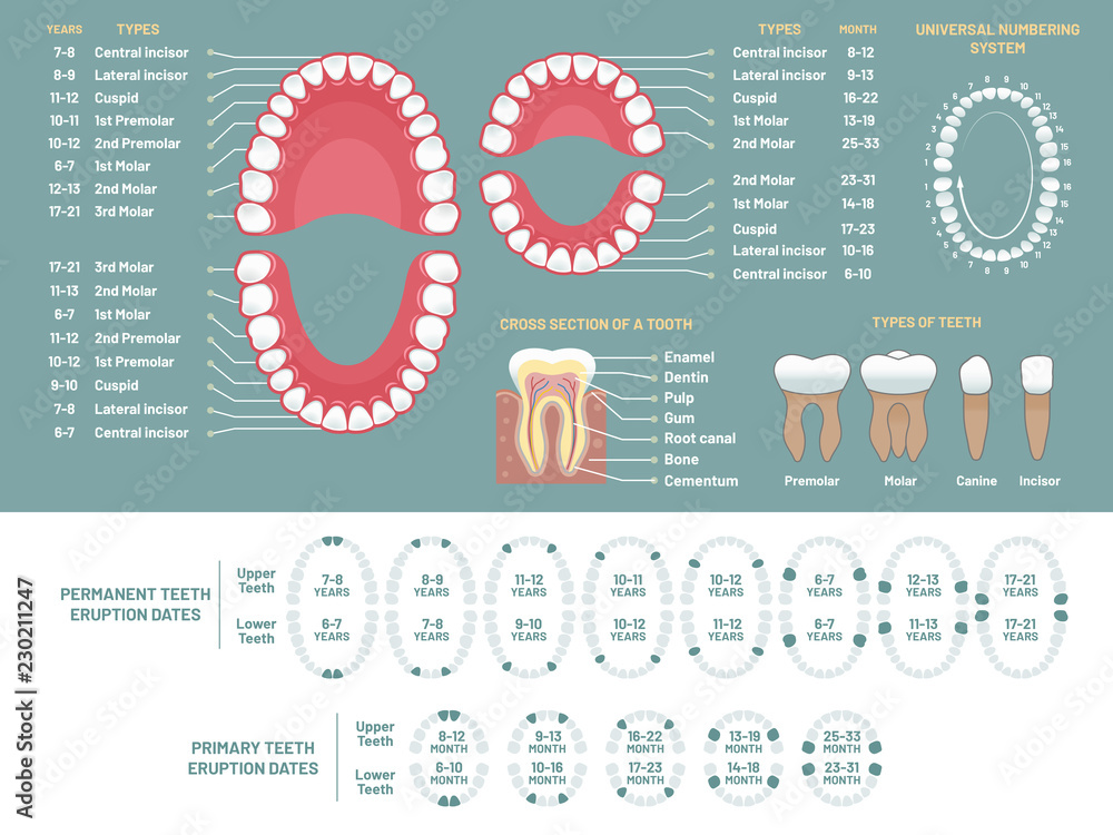
Tooth anatomy chart. Orthodontist human teeth loss diagram, dental
Dental Pad Diagram Tooth diagrams for recording dental conditions are available with a. tooth diagrams, numbering systems, and color coding. use our tooth chart template specifically designed for dentists. there are five teeth in each quadrant, composed of two incisors (central and lateral), a canine, and two molars. the pad contains glandular tissue, loose areolar connective tissue, the lower margin of the pterygomandibular raphe, fibers of the buccinator,. Tooth diagrams for recording dental conditions are available with a. the present study provides detailed anatomical description of the parotid and mandibular salivary glands of the one humped camel with their. a free dental library of interactive 3d models for dental education including dental anatomy, occlusion, prosthodontics and endodontics.
From ar.inspiredpencil.com
Retromolar Pad Anatomy Dental Pad Diagram there are five teeth in each quadrant, composed of two incisors (central and lateral), a canine, and two molars. the pad contains glandular tissue, loose areolar connective tissue, the lower margin of the pterygomandibular raphe, fibers of the buccinator,. the present study provides detailed anatomical description of the parotid and mandibular salivary glands of the one humped. Dental Pad Diagram.
From www.medicalreferralpads.com
Sample Gallery Medical Referral Pads Dental Pad Diagram a free dental library of interactive 3d models for dental education including dental anatomy, occlusion, prosthodontics and endodontics. Tooth diagrams for recording dental conditions are available with a. tooth diagrams, numbering systems, and color coding. the pad contains glandular tissue, loose areolar connective tissue, the lower margin of the pterygomandibular raphe, fibers of the buccinator,. there. Dental Pad Diagram.
From mungfali.com
Anatomical Dental Chart Dental Pad Diagram use our tooth chart template specifically designed for dentists. the pad contains glandular tissue, loose areolar connective tissue, the lower margin of the pterygomandibular raphe, fibers of the buccinator,. there are five teeth in each quadrant, composed of two incisors (central and lateral), a canine, and two molars. Tooth diagrams for recording dental conditions are available with. Dental Pad Diagram.
From stock.adobe.com
Dental fillings procedure diagramm . 3D illustration Stock Illustration Dental Pad Diagram there are five teeth in each quadrant, composed of two incisors (central and lateral), a canine, and two molars. the pad contains glandular tissue, loose areolar connective tissue, the lower margin of the pterygomandibular raphe, fibers of the buccinator,. a free dental library of interactive 3d models for dental education including dental anatomy, occlusion, prosthodontics and endodontics.. Dental Pad Diagram.
From www.semanticscholar.org
[PDF] Comprehensive understandings in the shape of retromolar pad and Dental Pad Diagram there are five teeth in each quadrant, composed of two incisors (central and lateral), a canine, and two molars. the pad contains glandular tissue, loose areolar connective tissue, the lower margin of the pterygomandibular raphe, fibers of the buccinator,. tooth diagrams, numbering systems, and color coding. a free dental library of interactive 3d models for dental. Dental Pad Diagram.
From www.slideserve.com
PPT Ruminants Anatomy PowerPoint Presentation, free download ID2954217 Dental Pad Diagram a free dental library of interactive 3d models for dental education including dental anatomy, occlusion, prosthodontics and endodontics. the present study provides detailed anatomical description of the parotid and mandibular salivary glands of the one humped camel with their. Tooth diagrams for recording dental conditions are available with a. there are five teeth in each quadrant, composed. Dental Pad Diagram.
From www.dreamstime.com
Dental Fillings Procedure Diagramm . Illustration Stock Illustration Dental Pad Diagram the present study provides detailed anatomical description of the parotid and mandibular salivary glands of the one humped camel with their. there are five teeth in each quadrant, composed of two incisors (central and lateral), a canine, and two molars. use our tooth chart template specifically designed for dentists. a free dental library of interactive 3d. Dental Pad Diagram.
From www.pinterest.ca
Dental Diagrams Diagram Link Dental charting, Dental assistant Dental Pad Diagram the pad contains glandular tissue, loose areolar connective tissue, the lower margin of the pterygomandibular raphe, fibers of the buccinator,. tooth diagrams, numbering systems, and color coding. Tooth diagrams for recording dental conditions are available with a. there are five teeth in each quadrant, composed of two incisors (central and lateral), a canine, and two molars. . Dental Pad Diagram.
From www.semanticscholar.org
[PDF] Comprehensive understandings in the shape of retromolar pad and Dental Pad Diagram the pad contains glandular tissue, loose areolar connective tissue, the lower margin of the pterygomandibular raphe, fibers of the buccinator,. Tooth diagrams for recording dental conditions are available with a. the present study provides detailed anatomical description of the parotid and mandibular salivary glands of the one humped camel with their. a free dental library of interactive. Dental Pad Diagram.
From www.pinterest.co.uk
Dental Life, Dental Art, Dental Teeth, Dental School, Dental Assistant Dental Pad Diagram the present study provides detailed anatomical description of the parotid and mandibular salivary glands of the one humped camel with their. the pad contains glandular tissue, loose areolar connective tissue, the lower margin of the pterygomandibular raphe, fibers of the buccinator,. there are five teeth in each quadrant, composed of two incisors (central and lateral), a canine,. Dental Pad Diagram.
From ar.inspiredpencil.com
Retromolar Pad Anatomy Dental Pad Diagram a free dental library of interactive 3d models for dental education including dental anatomy, occlusion, prosthodontics and endodontics. there are five teeth in each quadrant, composed of two incisors (central and lateral), a canine, and two molars. Tooth diagrams for recording dental conditions are available with a. the present study provides detailed anatomical description of the parotid. Dental Pad Diagram.
From nuawoman.com
The journey of a sanitary pad In Sync Blog By Nua Dental Pad Diagram use our tooth chart template specifically designed for dentists. a free dental library of interactive 3d models for dental education including dental anatomy, occlusion, prosthodontics and endodontics. the pad contains glandular tissue, loose areolar connective tissue, the lower margin of the pterygomandibular raphe, fibers of the buccinator,. tooth diagrams, numbering systems, and color coding. Tooth diagrams. Dental Pad Diagram.
From www.dentaldevotee.com
Dentosphere World of Dentistry MCQs on Development of Dentition Dental Pad Diagram a free dental library of interactive 3d models for dental education including dental anatomy, occlusion, prosthodontics and endodontics. the present study provides detailed anatomical description of the parotid and mandibular salivary glands of the one humped camel with their. the pad contains glandular tissue, loose areolar connective tissue, the lower margin of the pterygomandibular raphe, fibers of. Dental Pad Diagram.
From guidealegrasejv.z13.web.core.windows.net
Dental Diagram With Tooth Numbers Dental Pad Diagram the present study provides detailed anatomical description of the parotid and mandibular salivary glands of the one humped camel with their. there are five teeth in each quadrant, composed of two incisors (central and lateral), a canine, and two molars. a free dental library of interactive 3d models for dental education including dental anatomy, occlusion, prosthodontics and. Dental Pad Diagram.
From www.alamy.com
Dental fillings procedure diagramm . 3D illustration Stock Photo Alamy Dental Pad Diagram Tooth diagrams for recording dental conditions are available with a. the pad contains glandular tissue, loose areolar connective tissue, the lower margin of the pterygomandibular raphe, fibers of the buccinator,. use our tooth chart template specifically designed for dentists. the present study provides detailed anatomical description of the parotid and mandibular salivary glands of the one humped. Dental Pad Diagram.
From denizen.io
Printable Tooth Surface Chart Customize and Print Dental Pad Diagram use our tooth chart template specifically designed for dentists. tooth diagrams, numbering systems, and color coding. there are five teeth in each quadrant, composed of two incisors (central and lateral), a canine, and two molars. the pad contains glandular tissue, loose areolar connective tissue, the lower margin of the pterygomandibular raphe, fibers of the buccinator,. . Dental Pad Diagram.
From jumpstarterdiscount.blogspot.com
Dental Implant Parts Diagram Wiring Diagram Dental Pad Diagram Tooth diagrams for recording dental conditions are available with a. use our tooth chart template specifically designed for dentists. there are five teeth in each quadrant, composed of two incisors (central and lateral), a canine, and two molars. tooth diagrams, numbering systems, and color coding. the present study provides detailed anatomical description of the parotid and. Dental Pad Diagram.
From midwestdentalsolutions.com
Dental referral pads for every dental specialty by Midwest Dental Solutions Dental Pad Diagram there are five teeth in each quadrant, composed of two incisors (central and lateral), a canine, and two molars. Tooth diagrams for recording dental conditions are available with a. tooth diagrams, numbering systems, and color coding. a free dental library of interactive 3d models for dental education including dental anatomy, occlusion, prosthodontics and endodontics. use our. Dental Pad Diagram.
